Introducing our diagnostic equipment
A complete set of diagnostic equipment that supports a wide range of needs, from company medical checkups to comprehensive health screenings
We strive to detect illnesses in their early stages, using a full range of diagnostic equipment like the 3.0 Tesla MRI, 80-row CT, and 3D mammogram. Additionally, we have installed multiple endoscopy rooms to create more availability and ensure the easy scheduling of appointments.
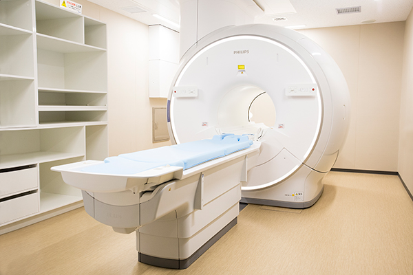
- 3.0 Tesla MRI
-
An MRI (Magnetic Resonance Imaging) is a diagnostic imaging device that can take cross-sectional images of any part of the human body using magnets and radio waves. It produces detailed images of the organs and blood vessels using magnetic forces as the patient enters a tube consisting of powerful magnets. A head MRI/MRA can screen for cerebral infarctions (including asymptomatic ones), aneurysms, and any thinning or narrowing of the blood vessels in the brain. A scan of the lower abdomen examines the prostate in men and the uterus and ovaries in women. The MRI can also be used to examine other body parts, such as the abdomen and spine. The advantage of the MRI is there is no concern for radiation exposure as it uses magnets instead of X-rays. Furthermore, it can produce cross-sectional images of the body and small blood vessels from all angles while lying down, without a contrast medium. We have several Philips Ingenia 3.0 T MRI scanners to meet a wide range of testing needs.
The advantage of MRI is that you do not have to worry about radiation exposure because you do not use X-rays but an examination using a magnet. It has features such as cross-sections at all angles without changing the position of the body just by sleeping, and obtaining blood vessel information without the use of contrast media. Nihonbashi Muromachi Mitsui Tower Midtown Clinic owns multiple PHILIPS 3.0 Tesla MRIs to meet various inspection needs.
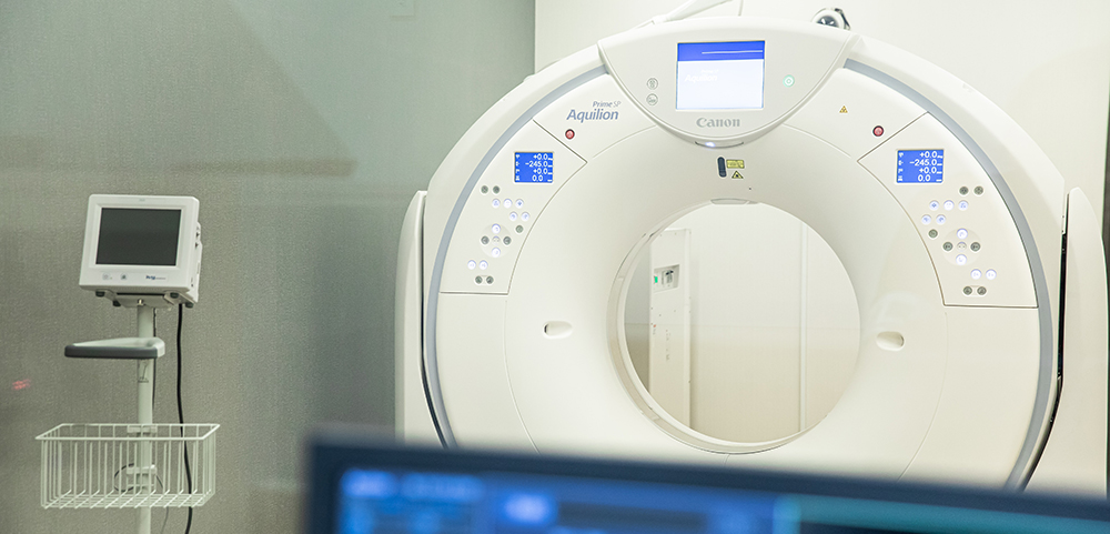
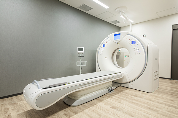
- 80-Row Multi-Slice CT
-
A CT scan uses X-rays to create cross-sectional images of the body. Using a multi-slice technology that can simultaneously capture 80 slices, the scanner can capture images of the head, chest, and abdomen, process them at high speed, and display them in real-time. The head CT can detect cerebral hemorrhaging, brain tumors, etc. The chest CT is useful in examining the lungs and mediastinum to screen for lung cancer. The abdominal CT can examine upper and lower abdominal organs (liver, pancreas, kidneys, etc.) and provide an even more detailed analysis when used with the ultrasound. The visceral fat CT measures the total area of abdominal visceral fat. It is useful in diagnosing metabolic syndrome. We use the Canon Aquilion PRIM (80 Row), a device that can produce higher quality images within a shorter timespan while also reducing the amount of radiation exposure. (※Radiation exposure is reduced by 60-70% compared to conventional models. )
Our comprehensive health screening includes a chest CT. The Midtown Course includes an additional upper and lower abdominal CT. You have the option of adding any CT scans to your original course.
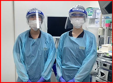
- Endoscopy room/Recovery room
-
We perform both upper and lower GI endoscopy procedures. Here we have multiple, highly coveted endoscopy rooms and make an effort to promptly guide patients through their checkups.
< You have the option of choosing between an oral or a nasal endoscope >
If you choose to have an upper GI endoscopy, you can do it through the nose or the mouth. With the nasal endoscopy, you can communicate with the attending physician during the exam because the camera is not obstructing your mouth. We recommend a nasal endoscopy for first-timers and those who have had a difficult experience with the oral endoscope. Our clinic aims to provide a painless experience, offering the option to use sedation so you can undergo the exam in a relaxing, semi-conscious state.
For those undergoing sedation, we must ask you to rest in our recovery room following the procedure.

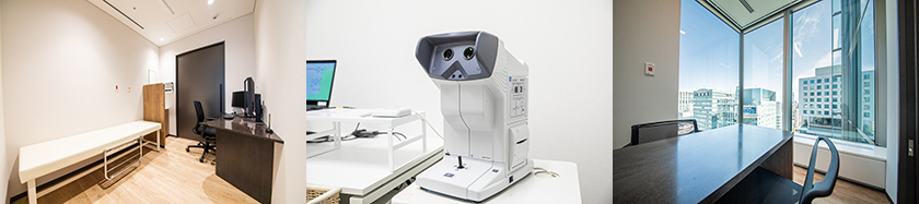
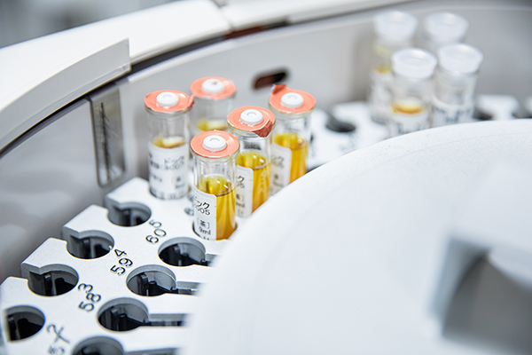
- On-site laboratory
-
The clinic has a laboratory built on-site, allowing us to provide prompt feedback on test results. Except for some blood tests such as the tumor markers, patients undergoing comprehensive health screening courses can have their basic blood panel analyzed during their appointment. You can immediately consult the physician regarding any concerns you may have.
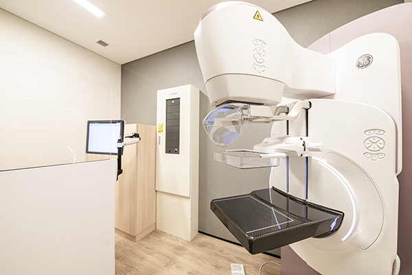
- Mammogram (2D/3D)
-
A mammogram is an X-ray picture of the breast. It is effective in detecting small lumps that cannot be found by palpation, as well as microcalcifications, which are early signs of breast cancer. The breast is evenly flattened out between two plates for imaging. Here we have a 3D mammogram (GE Healthcare Japan’s Senographe Pristina Mammography System), which allows a more detailed evaluation than the conventional 2D mammograms. The 3D mammogram takes images of the breast in layers, allowing tissue analysis using high-quality images, which is effective in the early detection of small cancers that are hidden in the mammary glands and are often challenging to find. Furthermore, at our clinic, all female-specific exams are performed and assisted by female staff, ensuring a safe and comfortable environment.
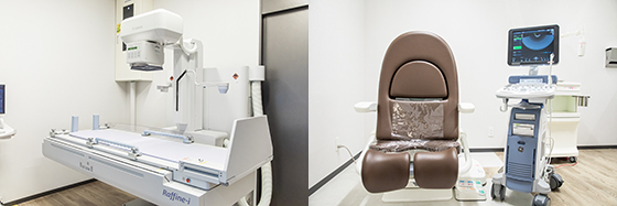
- X-ray equipment (Radiography)
-
Commonly referred to as an X-ray exam. X-rays are used to generate images of the chest, abdomen, bones, etc. The images are black and white, with material like bones that do not pass X-rays showing up white and body cavities that pass X-rays easily showing up black. The exam is routinely performed in comprehensive health screenings and medical checkups.
- Fluoroscopy equipment (Barium swallow)
-
A barium swallow examines the stomach using a barium-based contrast medium. We use a device equipped with a stopper function that prevents steep angling when the patient’s head is pointing down, a safety measure to avoid accidents, especially with older patients who may have difficulty supporting themselves.
- Ultrasound
-
During an ultrasound, sound waves are transmitted into the body, and the reflected waves are used to generate images of the insides. The abdominal ultrasound included in the comprehensive health screenings and medical checkups screens the upper abdominal organs like the liver, biliary tract, pancreas, kidneys, and spleen. We also perform breast, thyroid, heart, carotid artery, and transvaginal ultrasounds. The Canon Medical Systems’ fully digital ultrasound system allows more precise and accurate diagnostic testing than before.
- Electrocardiogram
-
The electrocardiogram records the electrical signals generated by the heart. The exam screens for arrhythmias while also evaluating the condition of the myocardium. You will lie on your back during the exam while six electrodes are attached to your chest. Another four are attached to each wrist and ankle. The entire exam lasts roughly a minute and incurs no pain.
- Ankle-brachial index (ABI)
-
The ABI test measures blood flow and screens for any hardening of the blood vessels or clogging. It can help monitor the progression of arteriosclerosis and risk of cardiovascular disease (stroke, myocardial infarction, etc.)
- Fundoscopy / Tonometry
-
A fundoscopy exam checks the blood vessels, retina, and optic nerves in the fundus through the lens of an ophthalmoscope. We use a non-mydriatic ophthalmoscope equipped with image processing capabilities for cataracts to capture more precise images of the fundus. It has auto functions such as high-speed autofocus and auto shot, which shortens the time spent in the exam room, reducing the burden on our patients. The tonometry exam primarily screens for glaucoma, measuring the hardness of the eye (intraocular pressure) by blowing a short puff of air or gently pressing a probe against the cornea. The exam incurs no pain and can be completed in one to two minutes.